MedicaBazaar

Registration

Language

Location

Sign In

Language And Payment

Sign In

Latest Searched
Laboratory Laboratory Medicine Other Laboratory Analysis Systems
Expanded Image
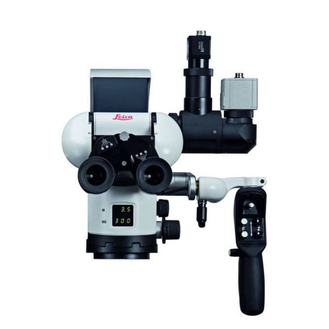
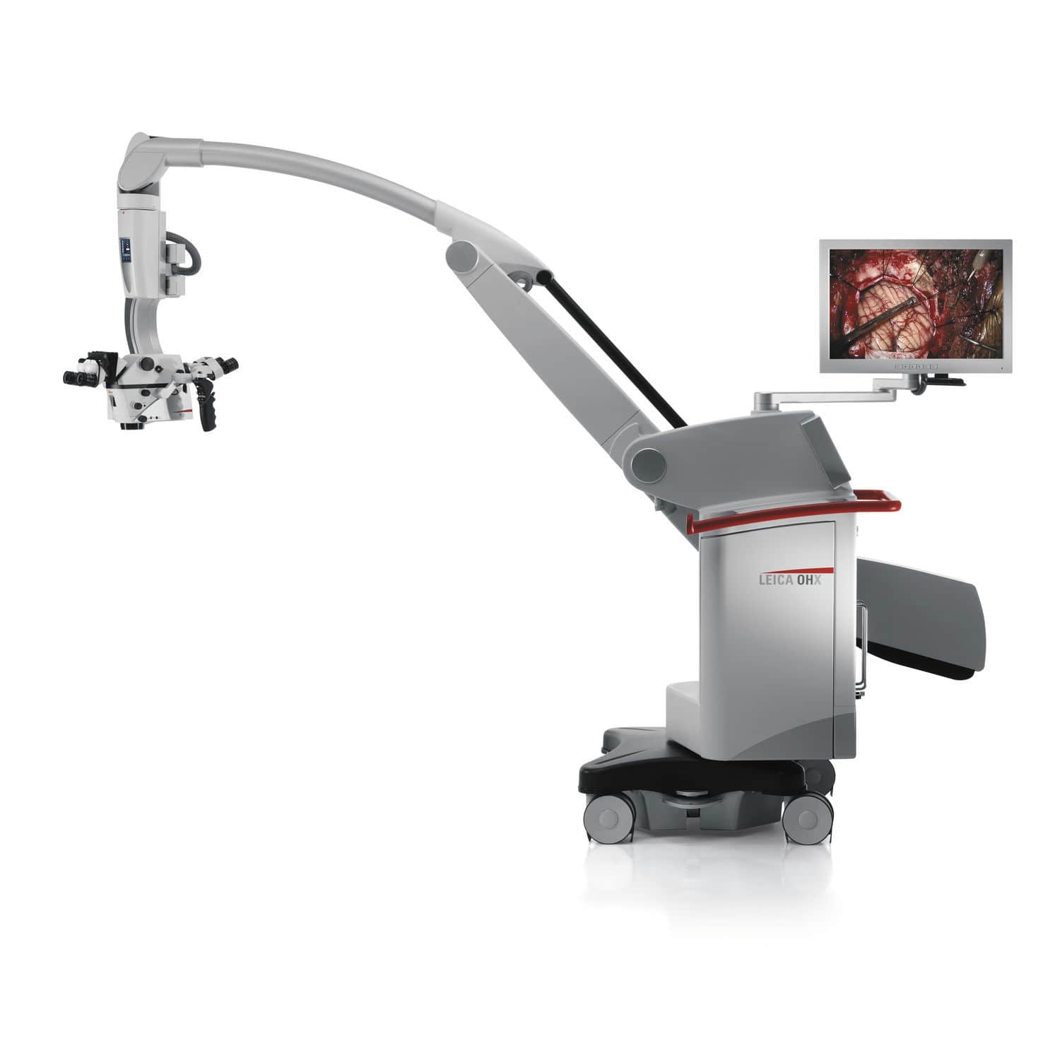
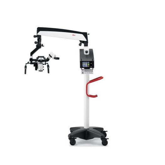
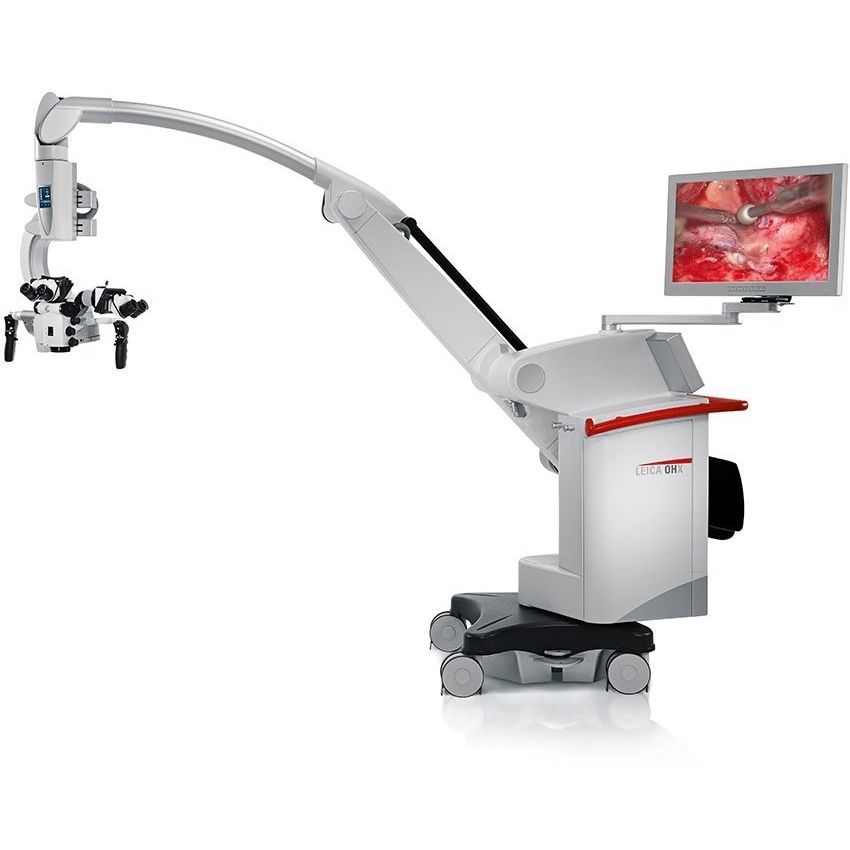
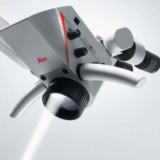
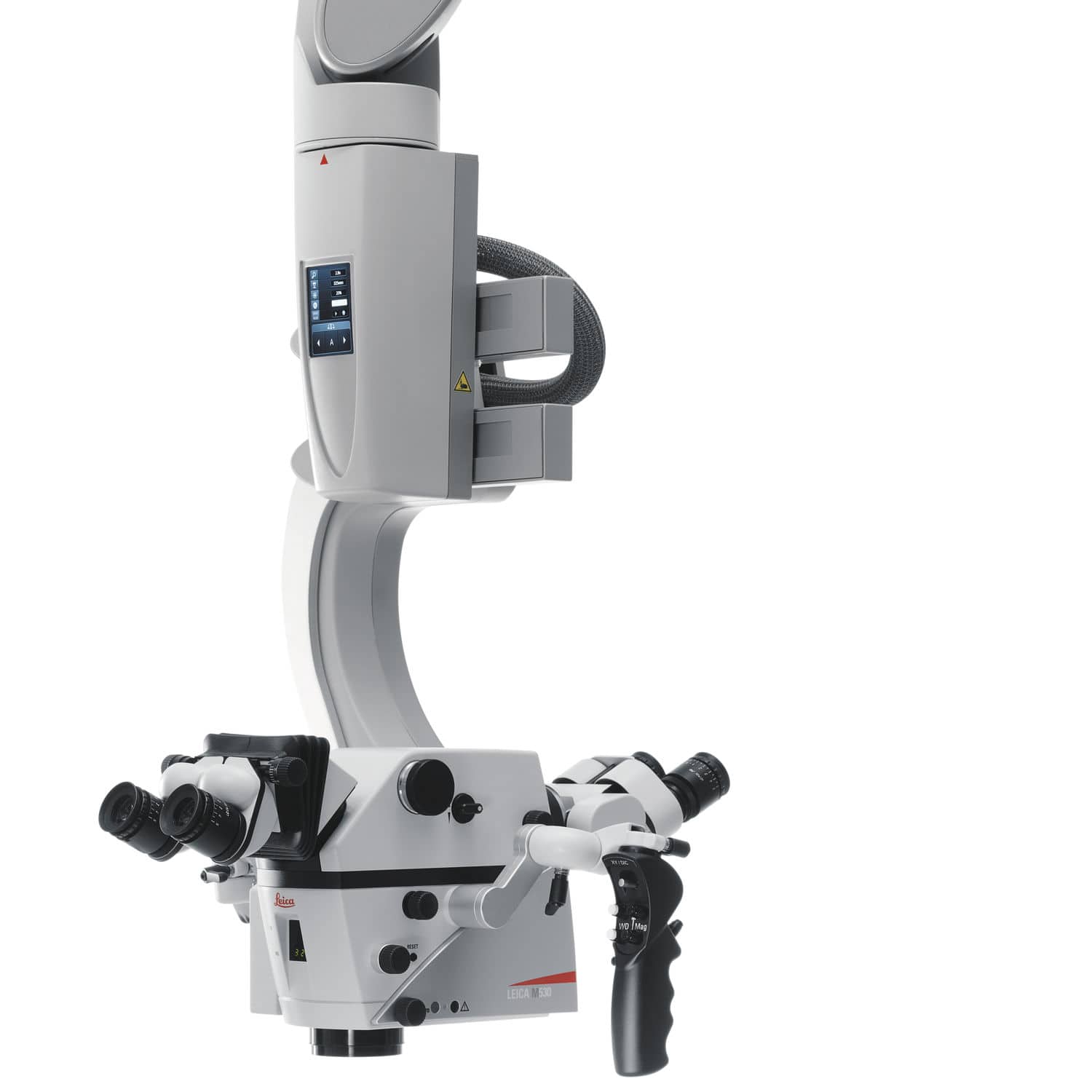
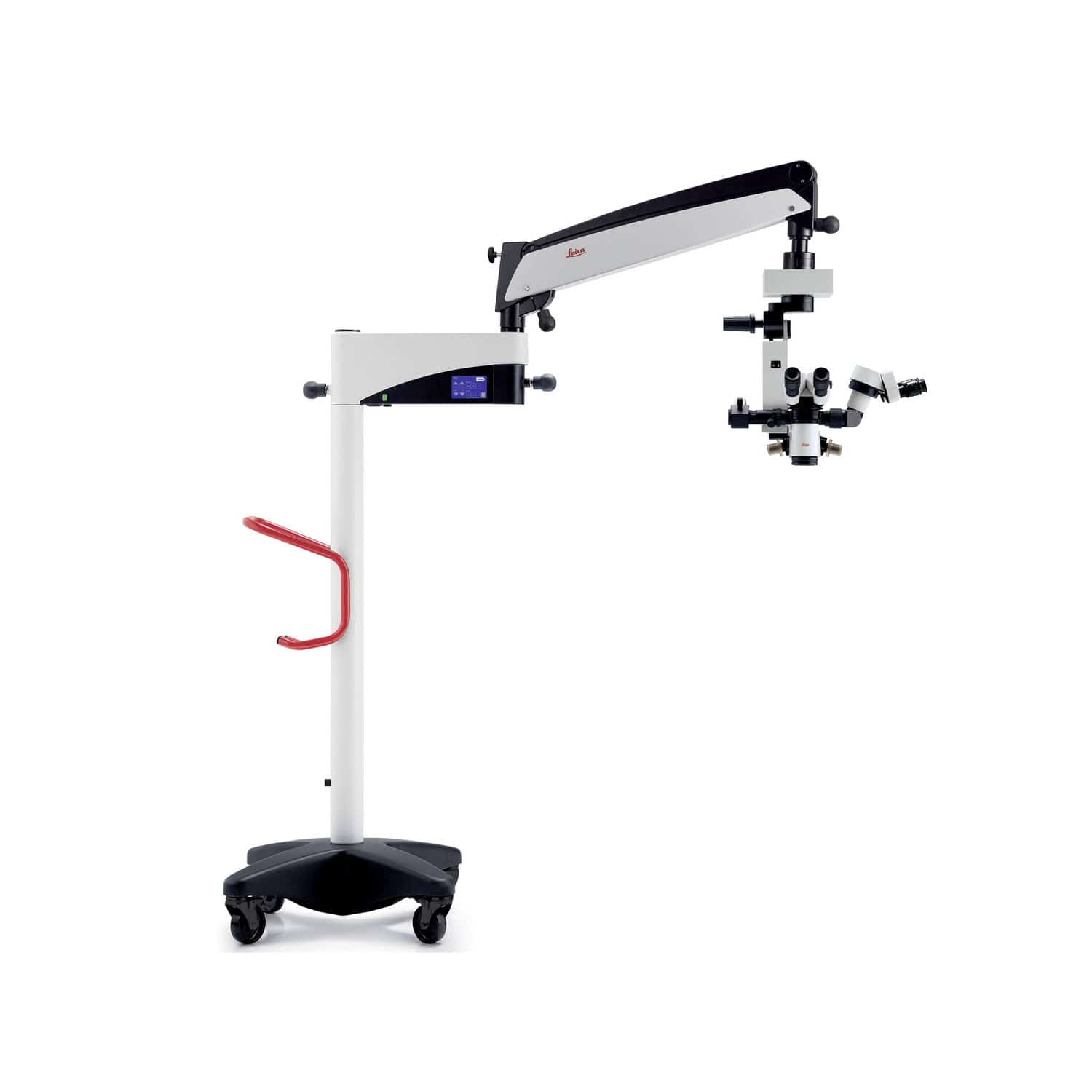
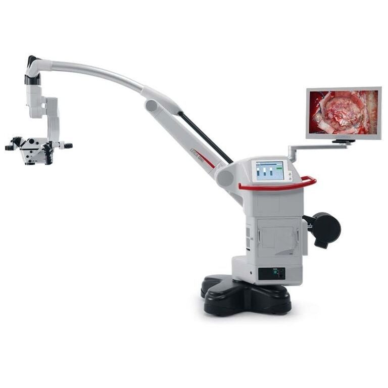
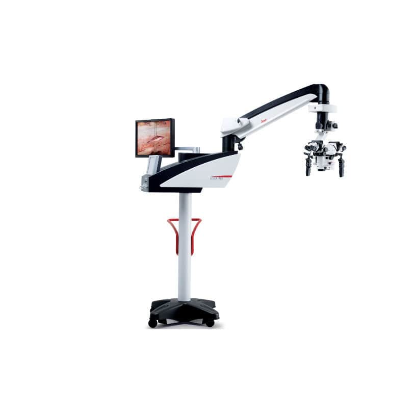
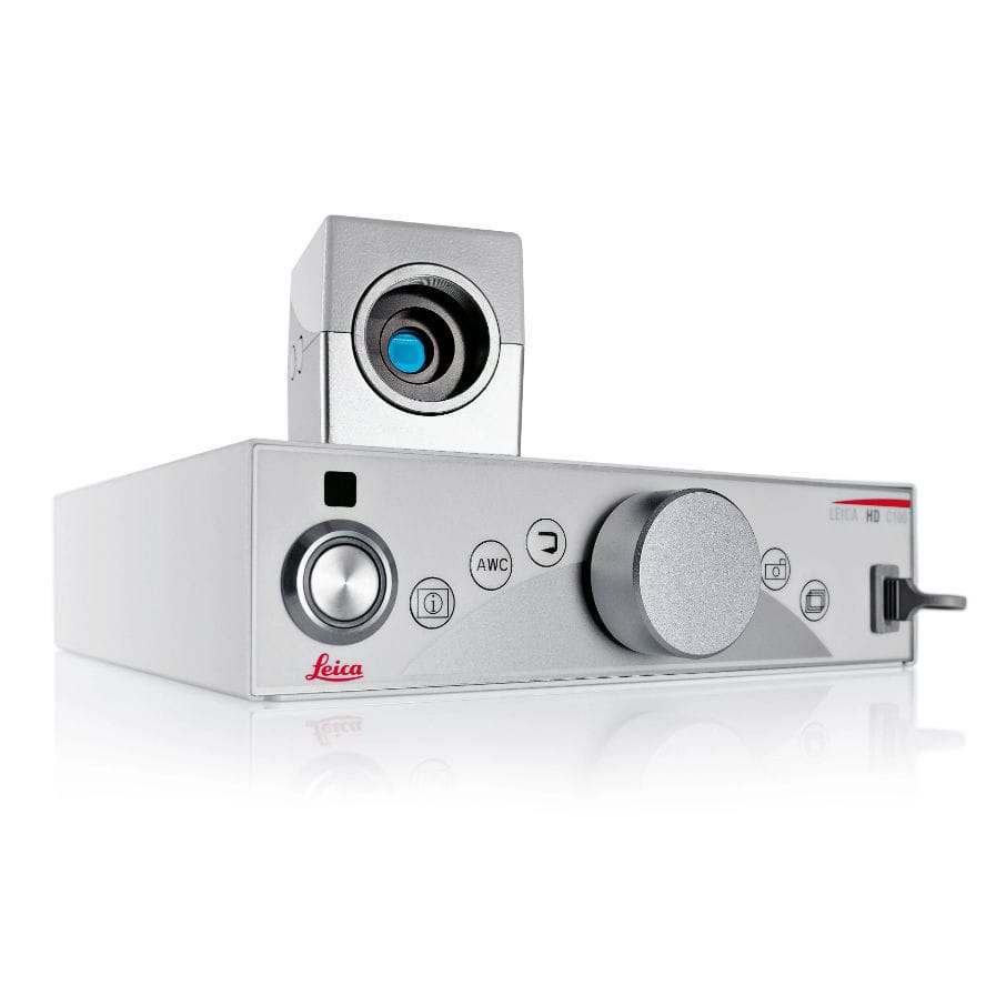
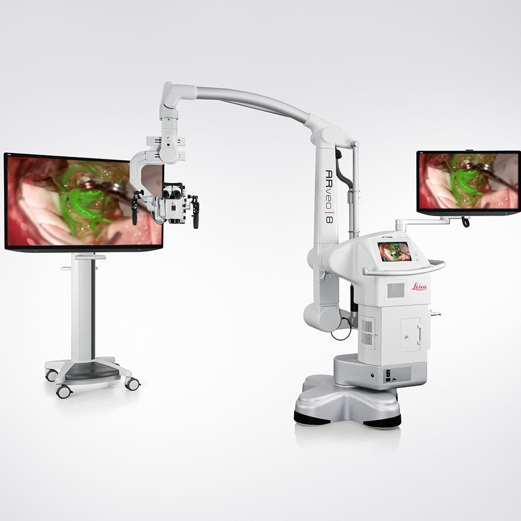
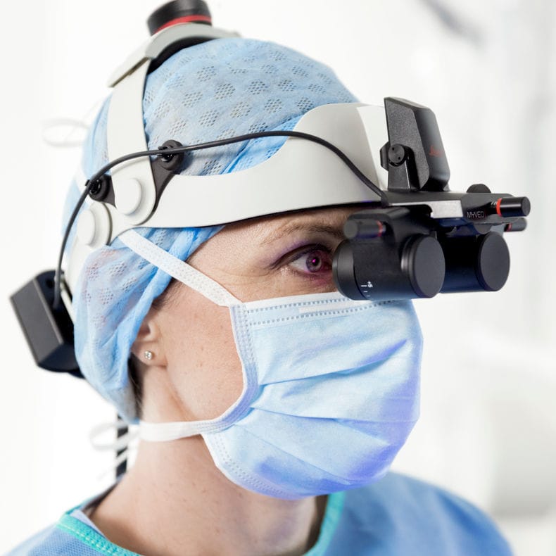
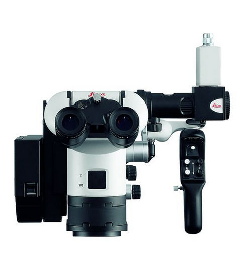
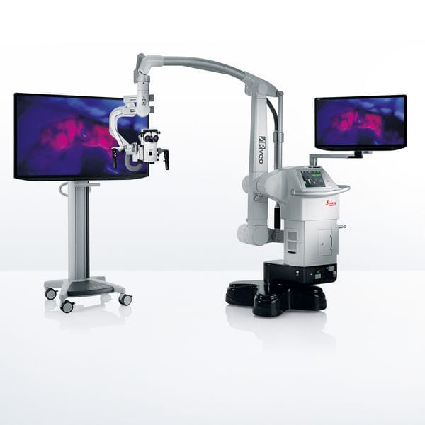
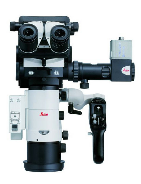
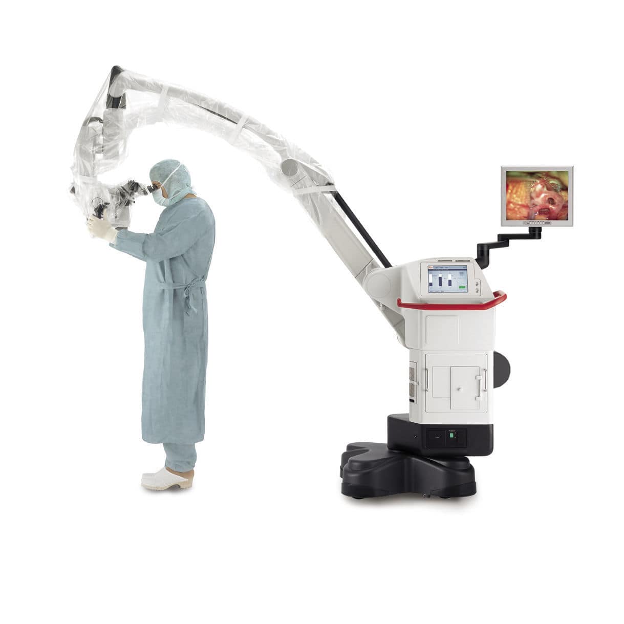
Operating microscope video camera
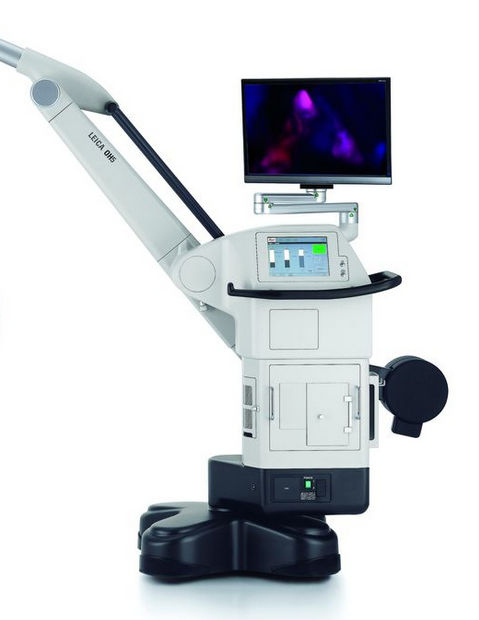
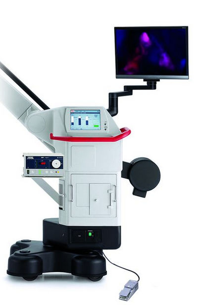
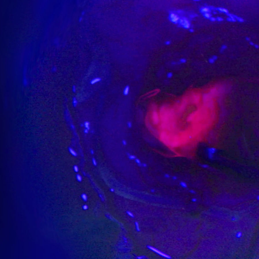
 FL400
Characteristics
Applications for operating microscopes
Light source fluorescent, white light
Resolution level HD
Technology digital
Description
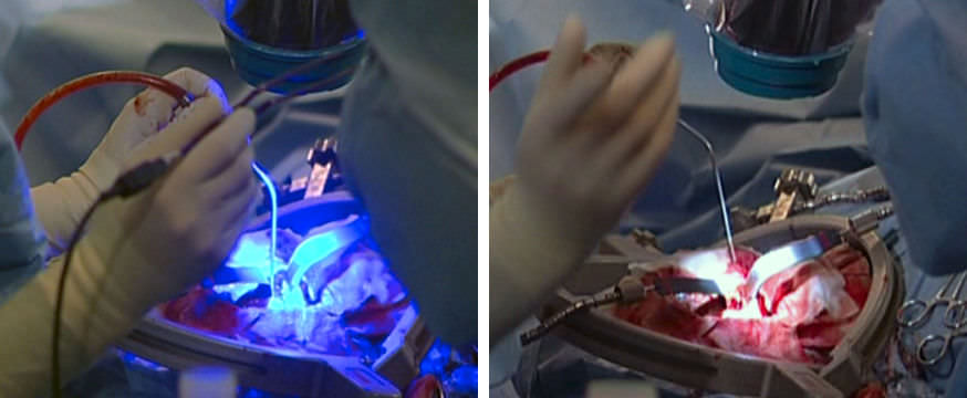
FL400 fluorescence with 5-ALA allows differentiation of tumor tissue from healthy brain tissue to support precise resection.

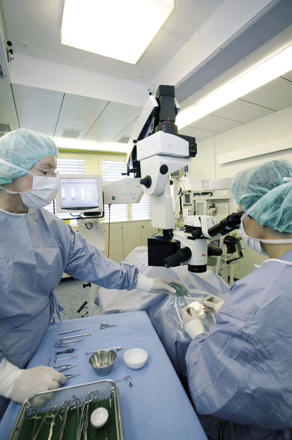
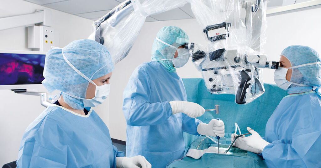
When resecting a malignant glioma, maximum removal of tumor cells with minimal impact to brain tissue is key to an optimal patient outcome. This is where FL400 fluorescence can provide support. In combination with the active substance 5 aminolevulinic acid (5-ALA), it helps distinguish both the bulk and margins of the tumor more easily. More visual information helps you to confidently perform a resection.

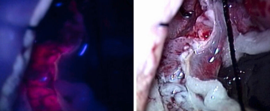
If it glows red, it's tumor
Under FL400 blue light mode, you and your assistant have a bright, high-contrast view of glioma cells. This aids with clear delineation of tumor margins for confident resection in real time.

How does it work?
Before surgery, the patient drinks a 5 aminolevulinic acid (5-ALA) solution. The 5-ALA is selectively taken up by glioma cells and converted into the fluorescent PPIX.
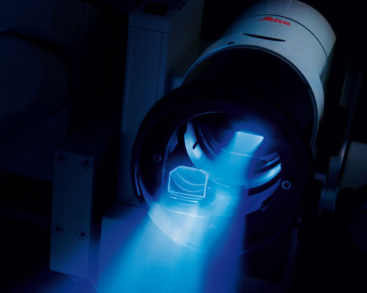
During surgery, you simply activate the FL400 module with a click of a button.
The FL400 module delivers intense, homogeneous blue excitation light, with a well-adjusted observation spectrum.
The PPIX in the tumor glows an intense pinkish red, enabling clear differentiation of tumor cells from healthy tissue

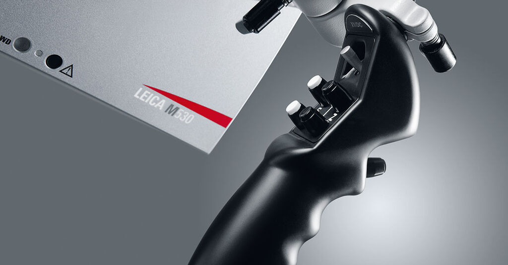
Easy handling for interruption-free work
Quickly and easily switch from white light to blue light fluorescence mode and back, or between fluorescence modes, for an interruption-free workflow.

As the FL400 module is fully integrated into your microscope, changing observation modes requires only the click of a button on the handgrip or footswitch.
Leica Microsystems
0 Followers
About the Company

Leica Microsystems develops and manufactures microscopes and scientific instruments for the analysis of microstructures and nanostructures.
Widely recognized for optical precision and innovative technology, the company is one of the market leaders in compound and stereo microscopy, digital microscopy, confocal laser scanning and super-resolution microscopy with related imaging systems, electron microscopy sample preparation, and surgical microscopy.


Dedication
Our work has made a difference for more than 160 years to scientists, engineers, surgeons, and even to humanity as a whole. No wonder we are so passionate about it. Coming from a scientific perspective, we always strive to deeply understand the work of our users in order to turn their needs and ideas into innovative solutions. This makes us proud partners of all those at the forefront of today’s innovation. A great responsibility that we meet with exceptional dedication – day in, day out.

Brilliance
Our users have brilliant minds. They expect the same brilliance from us as their partner. This entails a focus on state-of-the-art, top-quality products and excellent services based on a profound understanding of their respective fields of work. In other words: Our work must be as brilliant as the micro-images we represent.

Progress
We are all about progress. From research to product development to services, we continuously strive to improve on all levels to ensure that our users can always rely on the leading micro-imaging systems on the market. We help our users to advance their work every day and eventually, to accelerate the progress of humankind.
Products