Language And Payment
 USDEnglish
 TRYTurkish

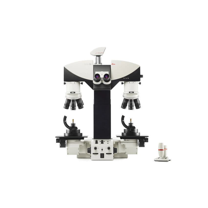
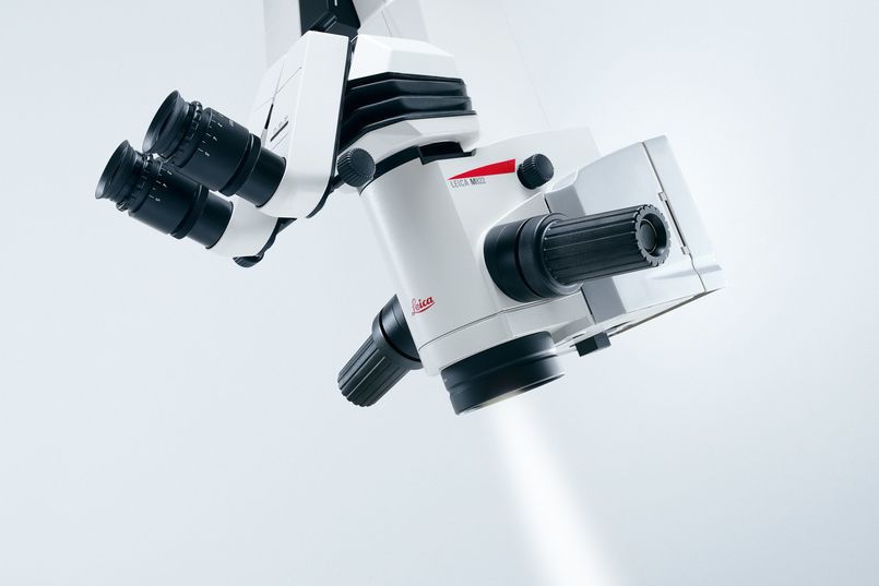
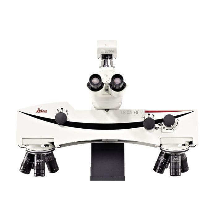
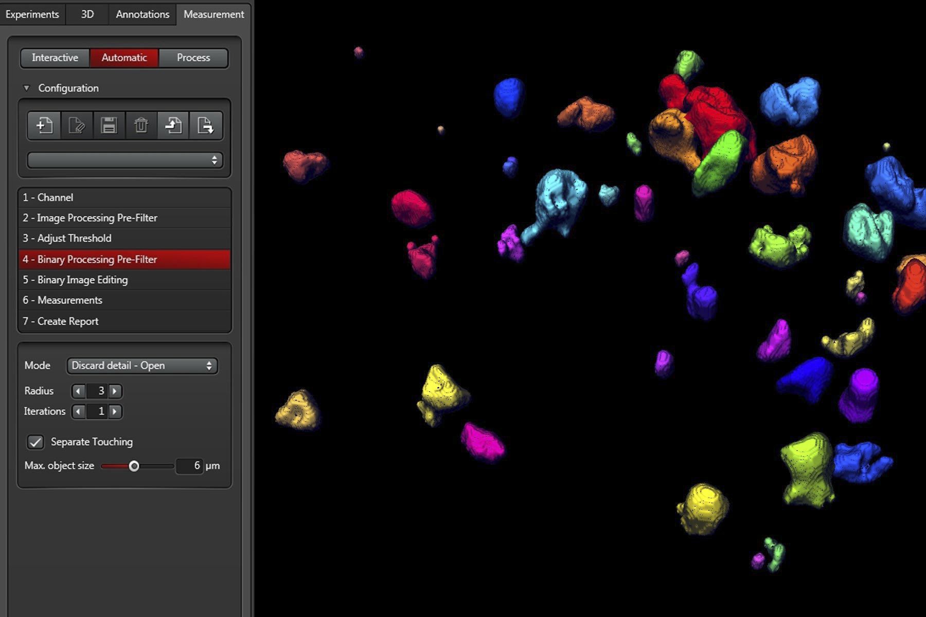
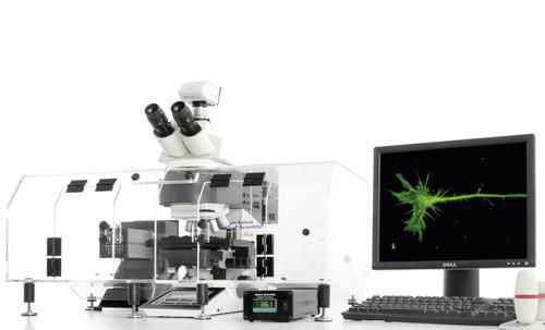
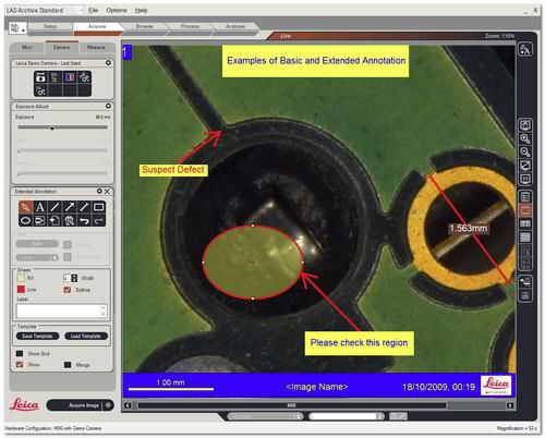
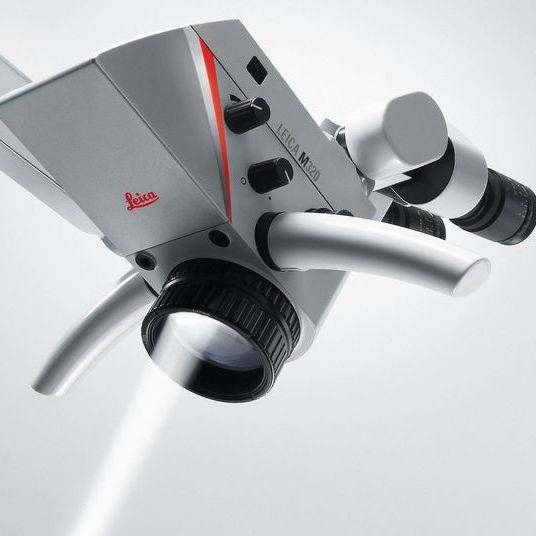
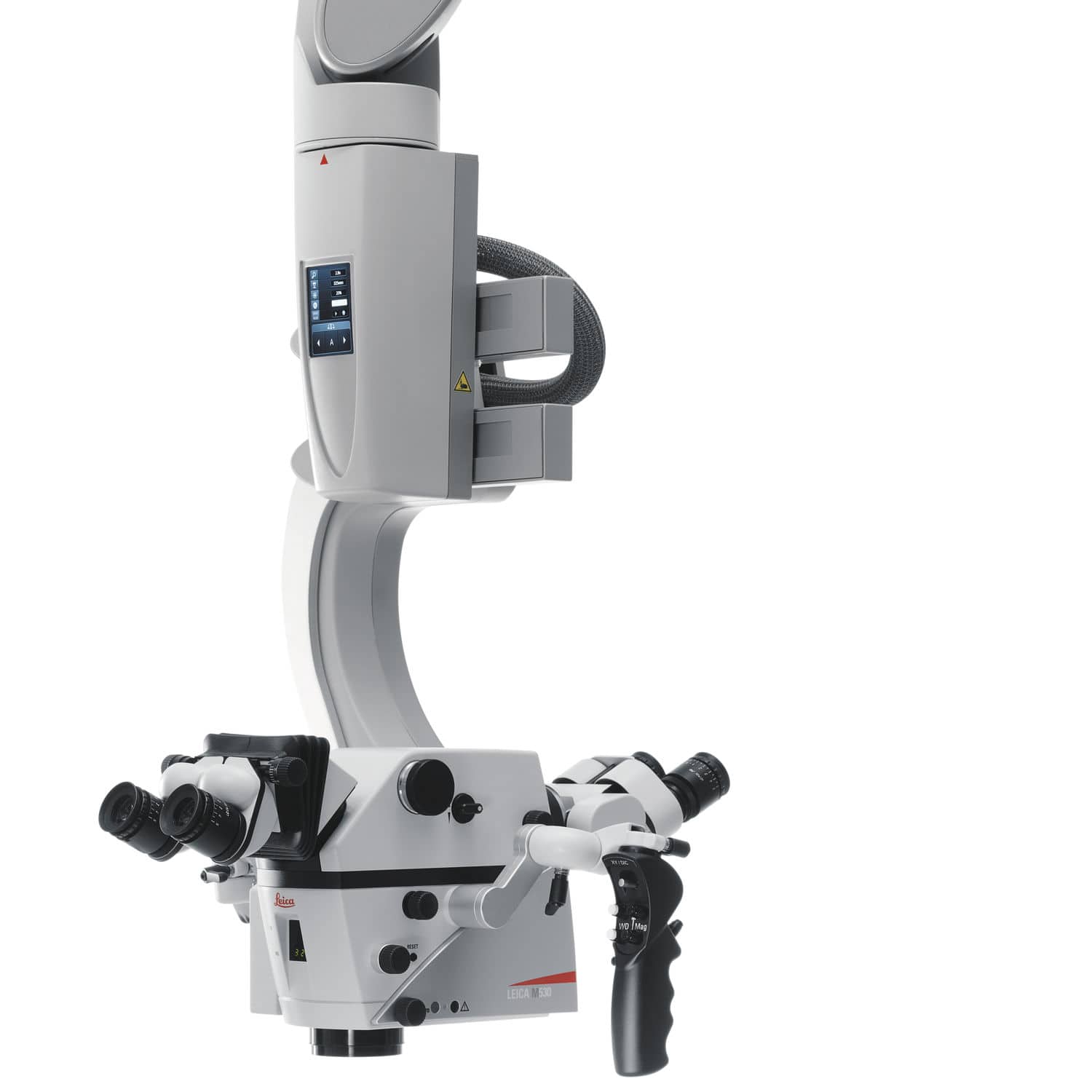
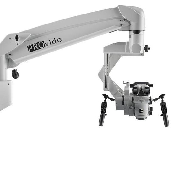
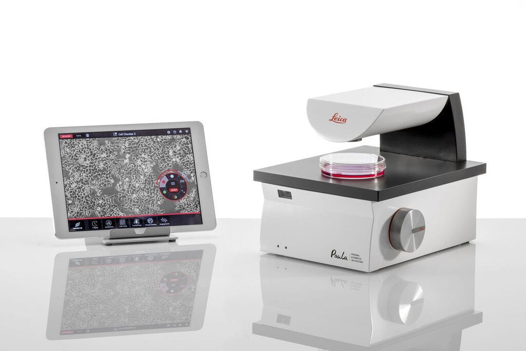
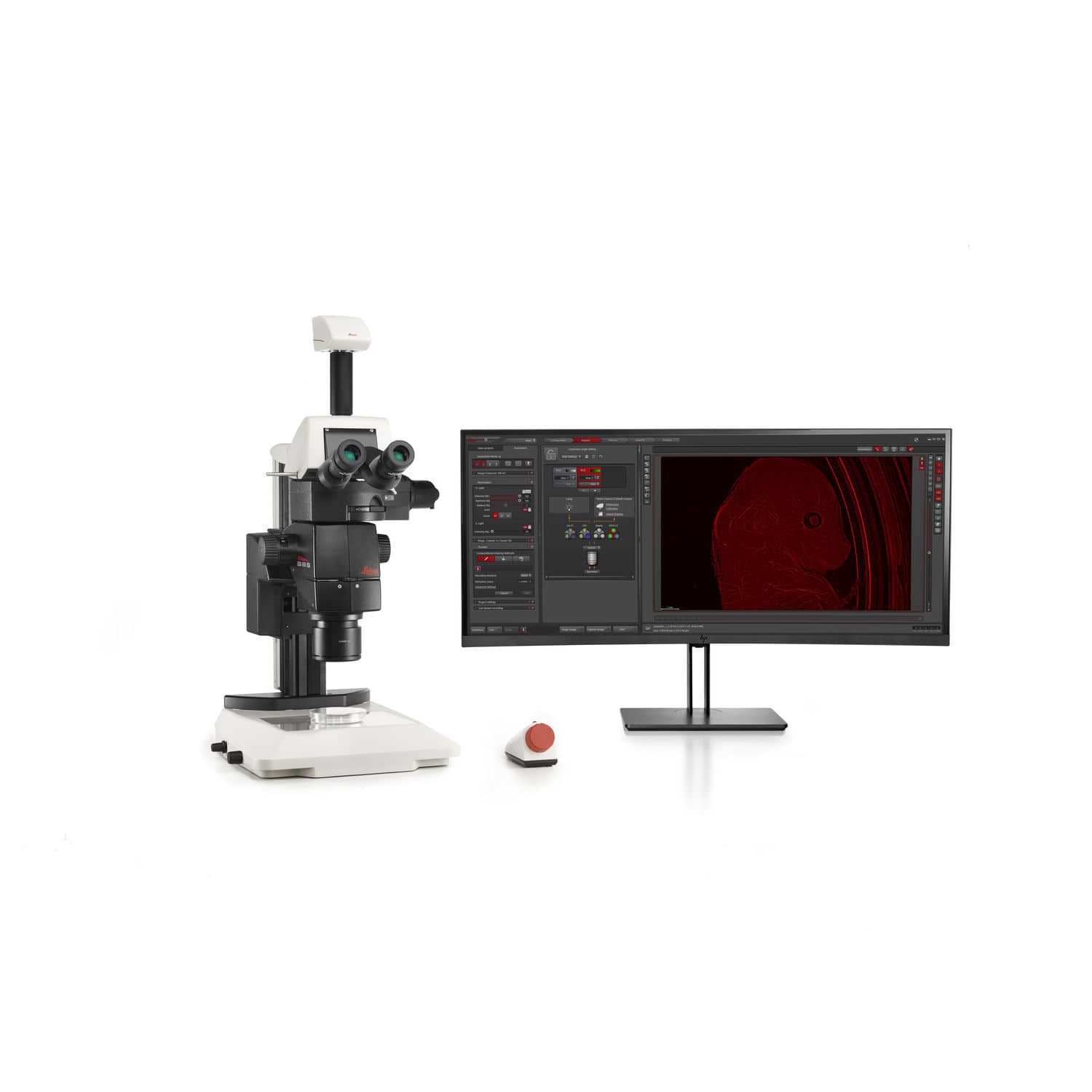
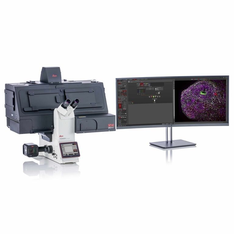
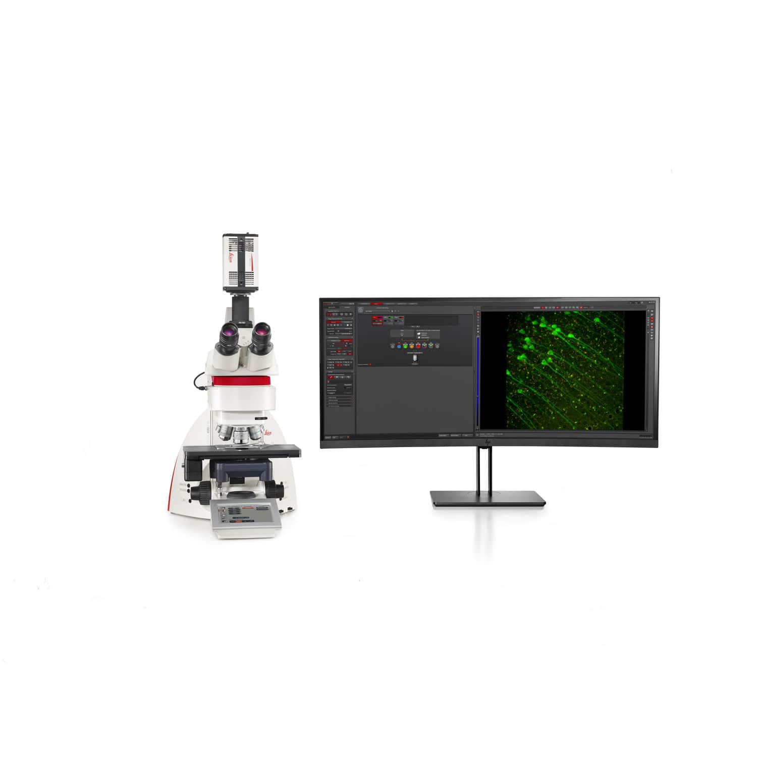
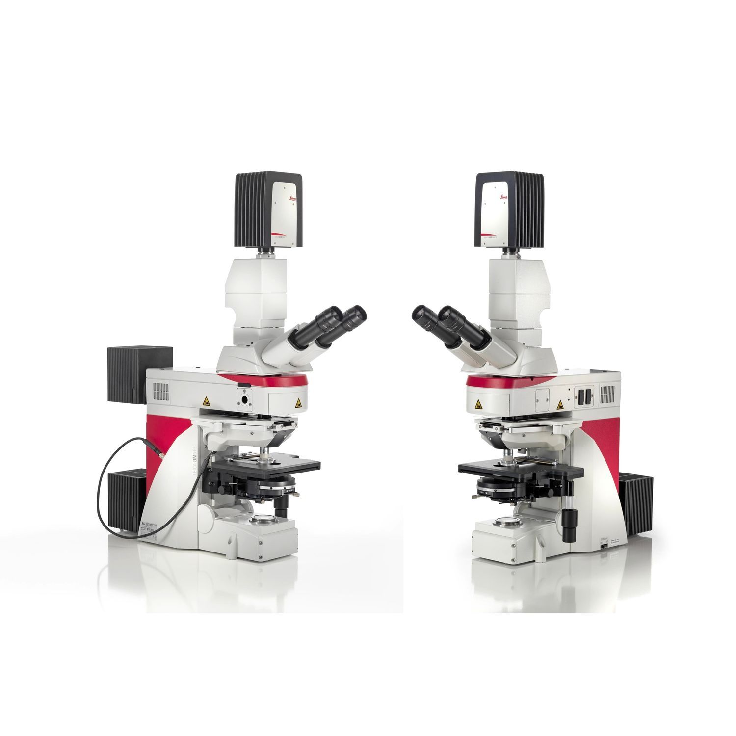
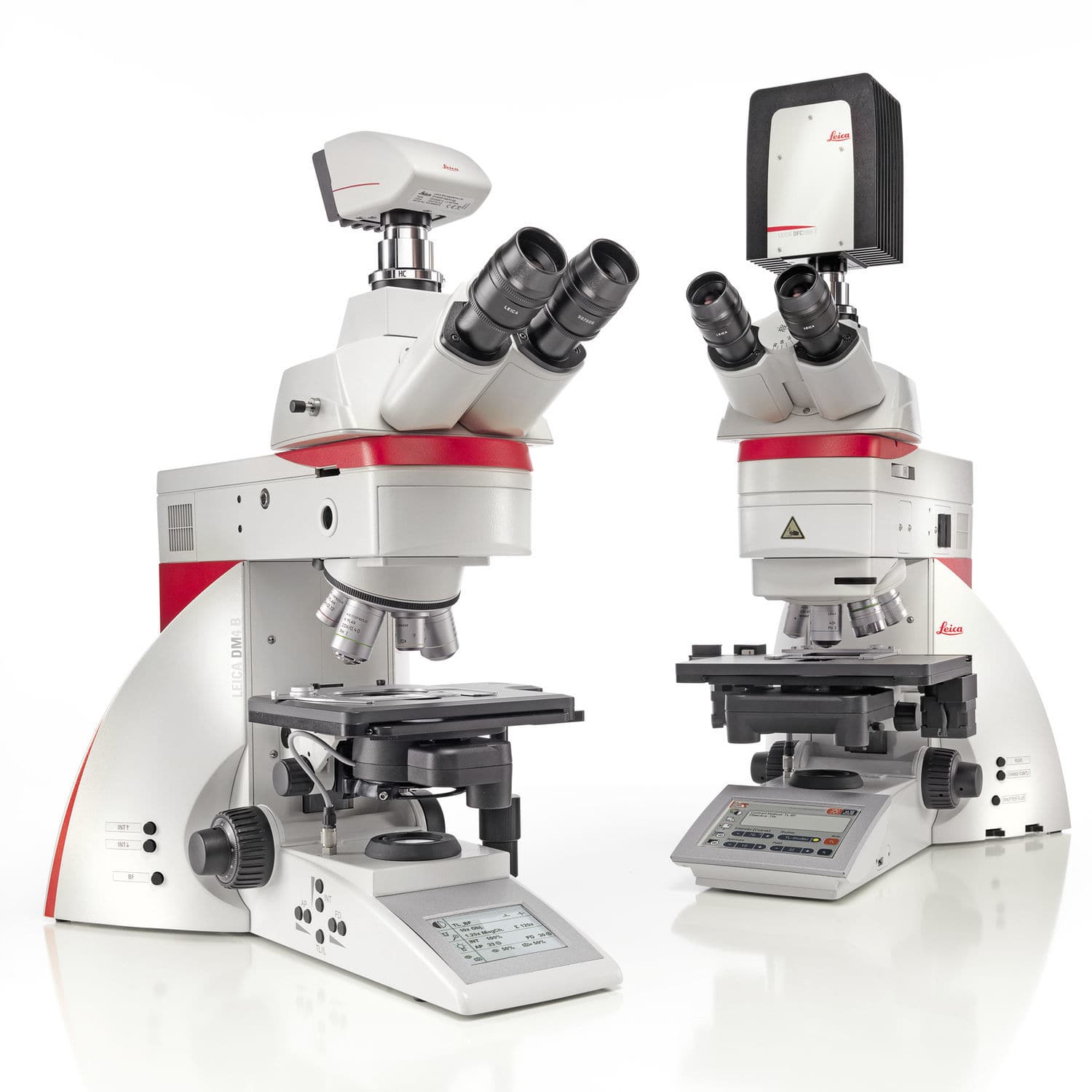
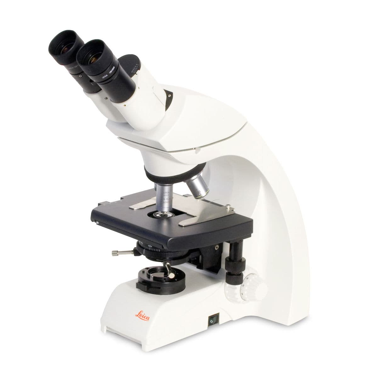
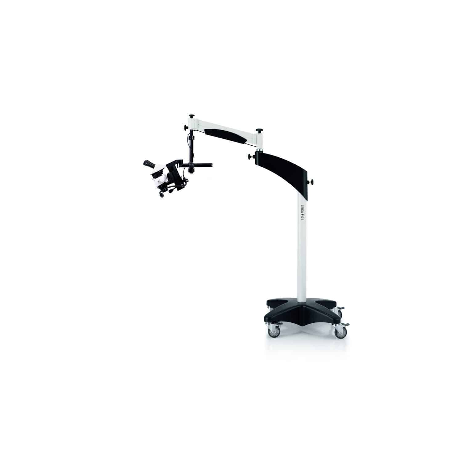
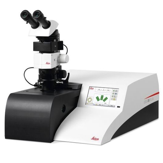
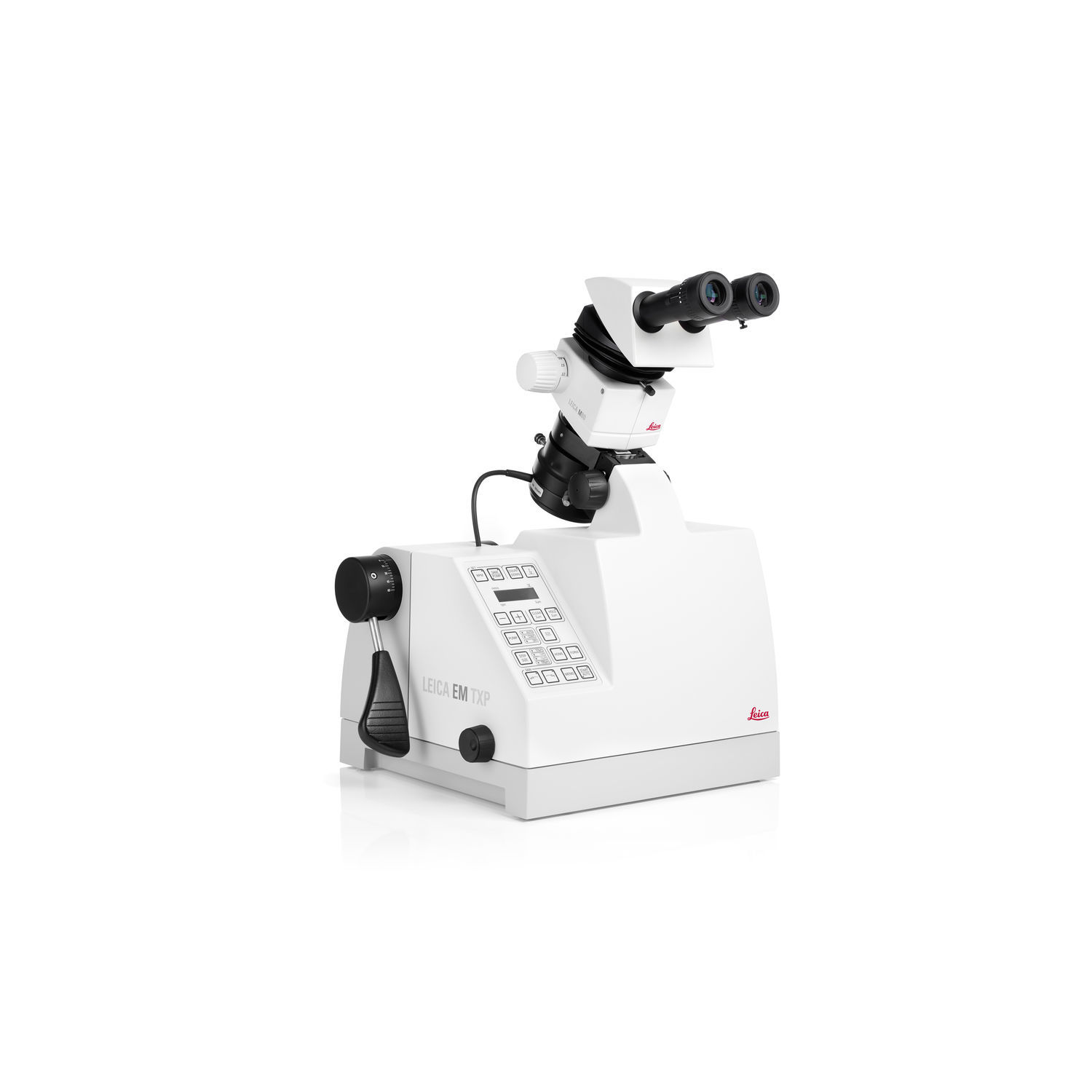
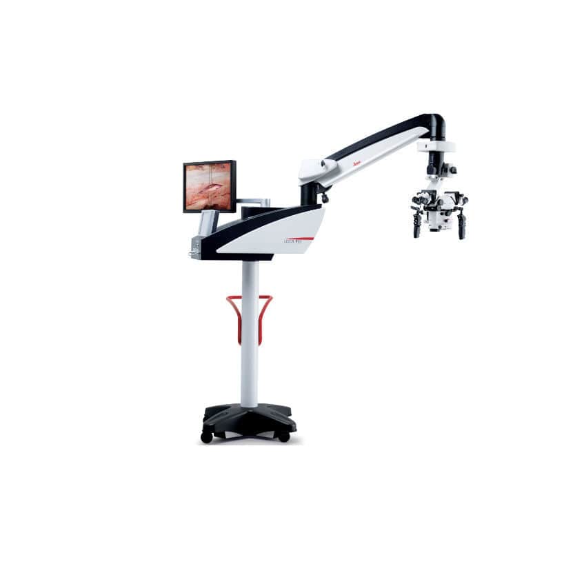
| applications | for life sciences applications, for biology, education, for research, for brain imaging, neuroscience |
|---|---|
| configuration | benchtop |
| ergonomics | upright |
| observation techniques | fluorescence, 3D, for live cells |
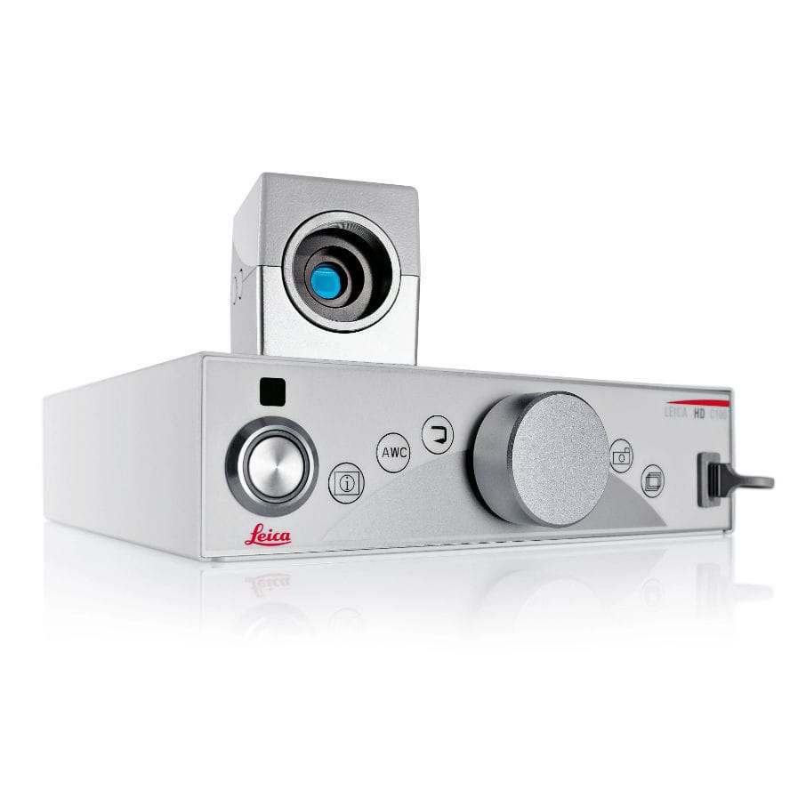
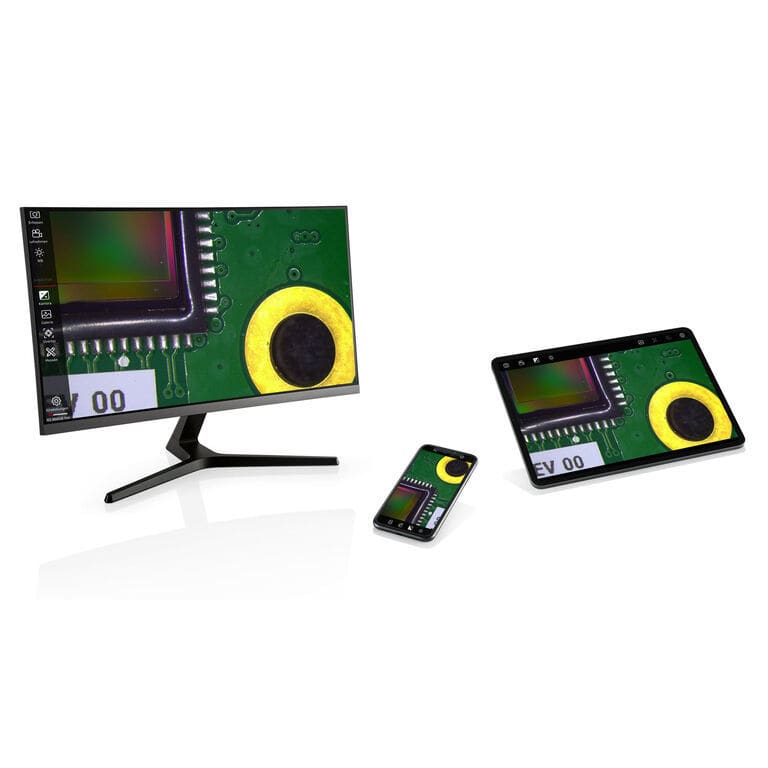
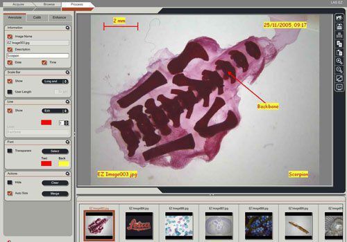
| options and accessories | with color camera, computer-assisted |
| other characteristics | high-resolution |
| type | optical |

 Request a Meeting